Inner ear hair cells, SEM - HD stock video
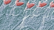
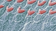
Inner ear hair cells. Coloured scanning electron micrograph (SEM) showing the sensory hair cells (blue) found in the Organ of Corti in the cochlea of the inner ear. These hairs are surrounded by a fluid called the endolymph. As sound enters the ear it causes waves to form in the endolymph, which in turn cause the hairs to move. The movement is converted into an electrical signal, which is passed to the brain. A single row of stereocilia can be seen at top, with three rows of outer hair cells below. Magnification: x3000 when printed at 10cm wide.


PURCHASE A LICENSE
Get personalized pricing by telling us when, where, and how you want to use this asset.
DETAILS
Credit:
51łÔąĎÍř #:
618321975
License type:
Rights-ready
Collection:
Image Bank Film
Max file size:
1920 x 1080 px - 484 MB
Clip length:
00:00:12:00
Upload date:
Release info:
No release required
Mastered to:
QuickTime 8-bit Photo-JPEG HD 1920x1080 25p
Categories:
- Cochlea - Inner Ear Videos,
- Biological Cell Videos,
- Inner Ear Videos,
- 10 Seconds or Greater Videos,
- Anatomy Videos,
- Art Product Videos,
- Biology Videos,
- Biomedical Illustration,
- Cilium Videos,
- Color Image Videos,
- Color Manipulation Videos,
- Digitally Generated Image Videos,
- Film - Moving Image Videos,
- Full Frame Videos,
- Green Color Videos,
- HD Format Videos,
- Healthy Lifestyle Videos,
- Horizontal Videos,
- Human Body Part Videos,
- Human Ear Videos,
- Human Hair Videos,
- Ideas Videos,
- In A Row Videos,
- Listening Videos,
- Magnification Videos,
- No People Videos,
- Organ of Corti Videos,
- Real Time Video Videos,
- SEM Videos,
- Science Videos,
- Sensory Perception Videos,
- Stereocilia Videos,
- Tilt Up Videos,